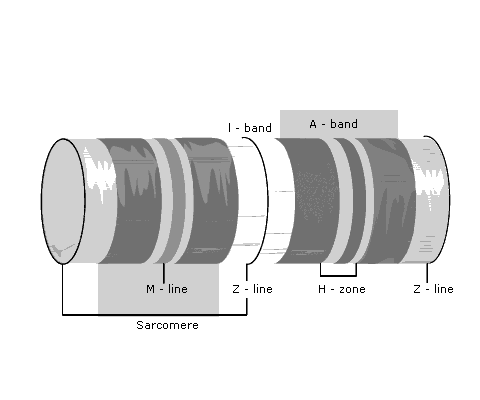
When viewed by polarized light, muscles have alternating dark and light zones (Fig 1). The regions that appear dark are called anisotropic, and the corresponding bands are known as A bands. The light regions do not refract and are said to be isotropic. These regions are called I bands.
Each I band is divided by a characteristic line known as a Z line. A sarcomere is the area between adjacent Z lines.
The darker A bands are made up of thick filaments, which contain the muscle protein myosin.
The thin filaments make up the I bands, and contain a long double helix arrangement of the protein actin (Fig 2).
Another protein, tropomyosin, is arranged in the grooves between the actin.
Molecules of troponin are located at intervals along the tropomyosin. Troponin T binds troponin to tropomyosin, troponin I inhibits the interaction of myosin with actin, and troponin C contains the binding sites for Ca2+.